PET Imaging (Positron Emission Tomography)
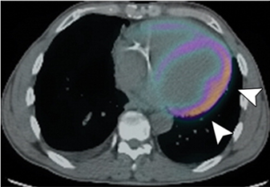
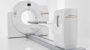
Pet uses radiopharmaceuticals, or tracers, to better understand the body’s chemistry, cell function, and location of disease. Unlike CT or MRI, which look at anatomy or body structure, PET studies look at body function. Different tracers are used for different studies. The tracer is absorbed by the area of interest and emits positrons, which are detected by the PET camera. A computer processes the pictures for the Radiologist or Cardiologist to interpret. PET imaging is safe and effective. The tracer is typically excreted out of the body through normal means.
PET procedures include:


