Nuclear Imaging
Nuclear Medicine uses radiopharmaceuticals, or tracers, to better understand how an organ is functioning. The tracer is usually given intravenously. Different types of tracers are used for different organs. The tracer is absorbed by the organ of interest and emits gamma rays, which are detected by the gamma camera. A computer processes the pictures for the Radiologist or Cardiologist to interpret. Nuclear medicine imaging is safe and effective. The tracer is typically excreted out of the body through normal means or simply loses the small amount of radioactivity that it originally had. Nuclear Medicine procedures include:
- Myocardial Perfusion Imaging
- Bone scans for infections, injuries, or tumors
- Hepatobiliary scans for liver and/or gall bladder function
- Gastric Empty scans for stomach function.
What are some common uses of the procedure?
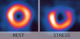
The most common cardiac nuclear medicine procedure enables the visualization of blood-flow patterns to the heart walls. It can be used to evaluate the results of bypass surgery or other procedures designed to restore the blood supply to the heart. Depending on the exact procedure performed, the myocardial perfusion scan takes between two and five hours.
How should patients prepare for the procedure?
Patients should avoid caffeinated and decaffeinated drinks and smoking for 12 hours before their examination. Patients should not eat or drink anything after midnight before the procedure. Comfortable walking shoes and loose-fitting clothes should be worn. Patients should tell the technologist and supervising physician if they have asthma or a chronic lung disease, or have problems with knees, hips, or maintaining balance.
What does the equipment look like?
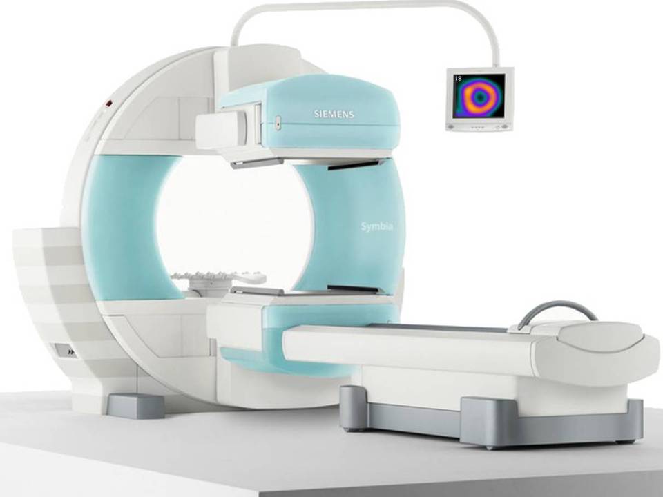
The imaging equipment, called a gamma camera, is similar to an x-ray camera. The detector portion of the camera can be changed to a variety of positions to obtain images of the body from different directions. A nearby computer console is used to develop the images of the heart.
What happens during the procedure?
Coronary arteries are evaluated by observing the changes in blood flow to the heart due to exercising. Patients undergo a stress test, either with exercise or drug infusion, to make their hearts work harder. The images obtained after exercise are compared with images of the heart obtained while resting. Comparison of the exercise and resting images is done to determine whether coronary blood flow has changed.
What are the benefits?
The test can be used to guide therapies designed to improved coronary blood flow and cardiac function. In addition, it can determine the ability of a patient to participate in an exercise program. Nuclear cardiac stress testing has been extensively studied and has been shown to be extremely safe, even in patients with known coronary artery disease.
What are the risks?
The test will be carried out under the supervision of a nurse or an advanced practice provider, with a cardiologist who is monitoring the test from outside the room. It is possible to experience chest pain or angina during stress test. The use of a radioactive substance will result in exposure to some radiation which is generally well tolerated.
A myocardial perfusion scan showing decreased blow flow to the heart muscle during stress conditions.


